In Part 1 of this article, we detailed the diagnosis and assessment of shoulder pain due to supraspinatous tendonitis. To summarize, this rotator cuff muscle is a common causes of shoulder pain and dysfunction.1 What is problematic for the acupuncturist is that the pain often refers to the deltoid region of the shoulder and sometimes distally down the arm and forearm. This deltoid region pain leads many practitioners to the diagnosis of large intestine (yang ming) and san jiao (shao yang) disorders, resulting in treatments that rarely suffice. Correct diagnosis is essential for a successful treatment outcome.
In TCM terms, injury to the supraspinatous muscle is most commonly diagnosed under the category "accident/trauma." The acute case usually has a definitive traumatic event. The chronic case is often a repetitive stress disorder due to the accumulation of microtrauma. The injury is at the level of the muscles and tendons, with qi and blood stagnation in the channels and collaterals. From a meridian (jing luo) perspective, the small intestine channel encompasses the belly of the supraspinatous (the region of SI 12); the Large Intestine channel covers the muscle-tendon junction and the tendinous aspect of the muscle (LI 16 to LI 15).
The following are several points and techniques to consider in the treatment of shoulder pain due to supraspinatous dysfunction. This protocol is organized into four steps; an approach that I use which makes point selection and needle technique simple, logical and systematic. It is both easy-to-understand and inclusive for acupuncturists from differing traditions and backgrounds.
STEP ONE
Use points and techniques that may have an immediate effect, such as decrease in pain or increase in range of motion.
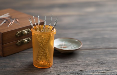
The Tendino-Muscle Meridians (SI 1, LI 1 Bleeding technique): Treatment to the jing-well points activates the tendino-muscle meridian. Try bleeding 10 drops of blood, which is often quite effective in treating shoulder pain. SI 1 treats the belly of the muscle (the region of SI 12), while LI 1 treats the area of the tendon and its attachment to the humerus (LI 16 to LI 15).
Opposite Side (contra-lateral): Many practitioners utilize treatment to ah shi points on the opposite shoulder. This technique should be considered, possibly combined with active movement of the affected shoulder.
Empirical Points (St 38): While the most effective use of St 38 is for a frozen shoulder, it may help with decreased range of motion sometimes associated with supraspinatous tendonitis.
STEP TWO
Use meridian and microsystem points not located at the site of injury. These are usually distal points chosen based on the signs and symptoms of the patient.
The Shu-Stream Point Combination (SI 3 affected side + Bl 65 opposite side): This right/left and upper/lower point combination is sometimes very effective in treating pain, and the shoulder is no exception. Remember, despite the patient reporting pain in the deltoid region of the shoulder, the primary stagnation is in the supraspinatous muscle. Thus, the small intestine (tai yang) channel is selected for treatment.
Traditional Point Categories (SI 6 xi-cleft point): SI 6 is one of the most effective points for acute cases of scapular shoulder pain. The practitioner, however, should not overlook other small intestine meridian points. Large Intestine (yang ming) points may be considered for pathology of the supraspinatous tendon (the region between LI 16 and LI 15). Palpate for sensitivity at LI 11 and points distal to the elbow.
Microsystems: These would include auricular therapy, and local (shoulder, master shoulder) and systemic points for pain (shen men, thalamus, adrenal, endocrine and muscle relaxation).
STEP THREE
Use points that benefit qi, blood and the zang-fu organs. Supraspinatous tendonitis is either an acute trauma or a chronic repetitive stress injury. Therefore, internal organ imbalances are usually not a factor in treatment. However, as with all cases of tendon pathology, consider liver imbalances.
STEP FOUR
Use local and adjacent points at the site of injury.
SI 12 (The supraspinatous muscle): This is located in the center of the suprascapular fossa, just superior to the spine of the scapula. This is also the precise location of both the trigger point and the motor point for the muscle.2 I usually treat SI 12 with two paired needles. Insertion is perpendicular, superior to the spine of the scapula. After insertion, angle the needle slightly inferior through the superficial muscle layers of the trapezius to the deeper and often taut portion of the supraspinatous. Look for a dense feeling in the needle. It may even "squeak" into this taut muscle tissue. Depth is from one-half to 1 inch, and if more than one needle is used, they may be spaced about 1 centimeter apart. The bony suprascapular fossa provides an end point to needle depth.
Keep the needle close to the spine of the scapula. Don't be lured into treating the tight bands of the upper trapezius muscle. In addition, patient position is very important to get the best access to the point. A seated patient, with arms adducted and shoulder depressed, works well. However, this increases risk for fainting and needle shock. The lateral recumbent (side-lying) position works equally well. I never treat this case with the patient prone (face down).
Electrical stimulation is very effective, but it also runs the risk of aggravating the condition. Consider the two needles inserted at SI 12, using electrical stimulation between them. Or combine SI 12 with a distal point.
LI 16 + LI 15 (The sub-acromial portion of the tendon): This is treating "above and below" (in anatomical terms medial and lateral to) the supraspinatous tendon that lies deep to the acromion. They are the most important points for both tendonitis and impingement syndrome. My needle technique is to insert LI 16 about .5 cun medial to its text location, and thread towards LI 15. The angle is from oblique to transverse. It may take several attempts and changes in angle to avoid hitting the bony clavicle or the spine of the scapula. Try to insert the needle from 1 to 1.5 inches, placed just superficial to the supraspinatous muscle. Make sure to avoid penetrating the pleural cavity. Use electrical stimulation between LI 16 and LI 15, preparing the patient for possible aggravation.
SI 11 (The infraspinatous muscle): The infraspinatous is often involved with supraspinatous dysfunction. The tendon of the supraspinatous has "extensions" which join the infraspinatous, thus both muscles are often involved in strain injuries.3 Consider SI 11 as a secondary point.
LI 14 (The deltoid muscle): This is an adjacent point at the insertion of the deltoid. It may be point-sensitive, as the deltoid muscle also assists in abduction and may be compensating, due to the distressed and weak supraspinatous. Consider LI 14 as a secondary point.
The prognosis for treatment of supraspinatous dysfunction varies and is directly related to the degree of structural damage to the muscle-tendon unit. Generally, consider treating twice a week for three weeks, then re-evaluate. Many cases without complications of tendon tears or sub-acromial bone spur have good results within six treatments. Some cases need treatment to continue at least once weekly after the first three-week period. The patient should be advised to discontinue any overhead and abduction movements of the arm that cause pain and aggravate the tendon.
Correctly diagnosing the supraspinatous as the site of primary stagnation of qi and blood is an important first step. The precise yet simple treatment outlined above usually results in very good treatment outcome. For practitioners unfamiliar with this protocol, hopefully you will approach this treatment with the enthusiasm and confidence.
Editor's Note: Portions of this article are excerpts from The Acupuncture Handbook of Sports Injuries and Pain. (Hidden Needle Press, 2009).
References
- Corrigan B, Maitland GD. Practical Orthopaedic Medicine. Butterworth-Heineman, 1989, p 41.
- Simons DG, Travell JG. Myofascial Pain and Dysfunction: The Trigger Point Manual, Vol. 1, (The Upper Extremities). Williams & Wilkins, 1983, pp 368-9.
- Dutton M. Orthopeadic Examination, Evaluation, and Intervention, Second Edition. McGraw-Hill, 2008, p 601.