One of the longest nerves in the body is known as the vagus nerve (VN). The VN is the 10th pair of cranial nerves that originates at the brain stem in the medulla oblongata. This nerve is part of the parasympathetic nervous system, which is a part of the ANS. Research suggests ear acupuncture can activate the VN.
A Deeper Look at the Parietal Lobe
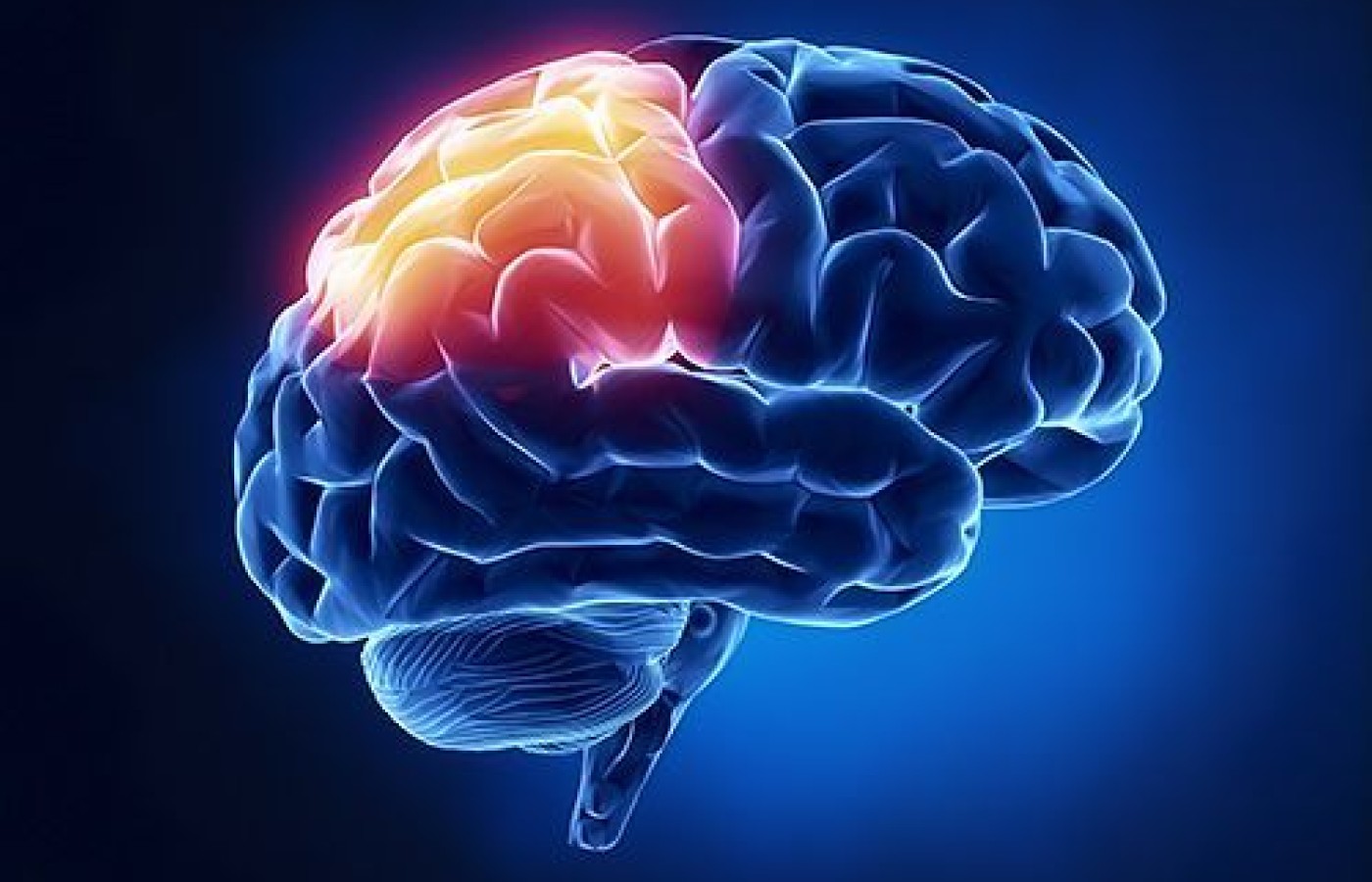
Many people associate the parietal lobe solely with the area of the brain that contains a sensory map of the body. However, it is much more complex in both structure and function. Let's explore both the anatomical structure and function, as well as their relevance in clinical practice.
Structure & Function
The parietal lobe contains several areas, but for the purpose of this discussion, let's focus on the primary somatosensory cortex (S1), secondary somatosensory cortex (S2), superior parietal lobe, inferior parietal lobe, and intraparietal lobe.
The primary somatosensory cortex (S1) provides touch, vibration, joint position sense, and two-point discrimination. The secondary somatosensory cortex (S2) receives information from S1 on pain and temperature. For this reason, the sensory line in scalp acupuncture is used often for somatic pain disorders.
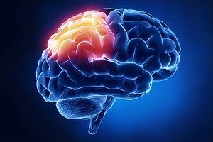
The superior parietal lobe plays a role in spatial awareness. Damage from stroke, for example, can cause hemispatial neglect. The inferior parietal lobe plays a role in both spatial awareness and body image, and damage to this area can cause different types of apraxias, which can be of movement, but also of speech.
An important consideration when discussing functions of the parietal lobe is that they differ slightly from left to right. The left inferior parietal cortex, specifically the angular gyrus, is involved in word comprehension – both written and spoken. Sentence comprehension and conceptual processing of verbal and written information involves this region of the brain that lives in close proximity to Wernicke's area, another key region in speech comprehension. Brain imaging studies show extensive activity in the left angular gyrus during the planning and execution of writing.
Following concussions and traumatic brain injuries, people commonly report difficulty with speech comprehension and reading comprehension, difficulty putting thoughts into fluent sentences, "word salad" (mixing up words in a sentence structure or say words they didn't mean to say), and often notice a decline in handwriting.
The parietal lobe is also highly integrated with vision and eye movements. The areas of the parietal lobe involved in visual pathways are the dorsal visual pathway and intraparietal areas.
The dorsal visual pathway is an important aspect of parietal lobe function that allows the brain to process "where" objects are in space. Smooth pursuit eye movements that track where targets are moving rely on parietal lobe function. The intraparietal area is involved in various types of eye movements and visual processing. Strokes and brain injuries affecting the parietal lobe can result in eye movement disorders.
Key Questions to Ask Your Patient During Intake
Given the functions of the parietal lobe, there are several key questions that can be used during an intake to suggest further examination of the parietal lobe would be valuable:
- Do you have chronic pain?
- Do you have trouble following driving directions?
- Are you frequently getting lost driving or walking?
- Do you have trouble finding your parked car?
- Do you often get right and left mixed up, or have to think about it harder than seems normal?
- Are you having trouble comprehending what you hear and read?
- Has your body image changed?
When working with people with chronic pain, it is important to appreciate that chronic pain lives in the brain and involves complex circuits that include the parietal lobe, amygdala, anterior cingulate gyrus, and hippocampus. Identifying poor parietal lobe function by assessing not only the somatosensory cortex, but also the superior, inferior and intraparietal regions while distinguishing whether the deficit is in the right, left or both lobes, can provide important information to direct acupuncture treatment strategies.
Functional Examinations
There are some simple bedside tests that can be done to assess the parietal lobes. Remember, the left parietal lobe contains a map of the right side of the body, and the right parietal lobe contains a map of the left side of the body. The right parietal lobe also has a redundant map of the right side of the body; however, for the purpose of assessment, we focus on the contralateral aspect.
When performing functional exams, it is essential that we never "hang our hat" on one single finding. This is why a combination of history, intake and exam should all add up to a meaningful understanding of the issue. For this reason, we will cover several different parietal lobe tests.
Toe Identification: With the patient laying supine, eyes closed, touch a toe and ask them to identify which toe is being touched. Move back and forth between feet, testing different toes and even coming back to ones incorrectly identified. Document the percentage of accuracy.
Next, perform a quick joint position sense test. Move the 2nd toe either toward the patient or away from the patient, and have them tell you which direction they sense the toe moving.
Palm "Reading": A graphesthesia test is another good parietal lobe test. Take the sharp end of a reflex hammer and with the patient's eyes closed, draw capital letters on the palm of the hand (tell them the letters will be facing you) and have them identify which letters are being drawn. Do 10 letters on each hand and document the accuracy.
Tracking the Thumb: Due to the parietal lobe's involvement in smooth pursuit eye movements, we can use slow pursuits as another functional way to assess the parietal lobe. Assess rightward pursuits separate from leftward pursuits. Have the patient follow your thumb from center to the right (go approximately 18 inches laterally) several times, and then do the same going to the left.
Look for differences in the smoothness of the movement. Are the eyes staying on the target, or do they look like they are skipping to keep up? Rightward pursuits provide an evaluation of the right parietal lobe; leftward pursuits provide an evaluation of the left parietal lobe.
How Does All of This Information Add Up?
Let's say you have a patient with a history of multiple concussions, and their main complaint coming into your clinic is chronic right shoulder pain and neck pain. If you observe that this person gets 100 percent correct on the left for toe identification and 50 percent correct on the right; 100 percent correct on graphesthesia testing on the left and 60 percent correct on the right; and their slow pursuit eye movements are more jerky going right, what do you know?
These findings would indicate poor functioning of the left parietal lobe. This deficit may include the region of the left somatosensory cortex that contains a map of the right shoulder. This may be contributing to chronic right shoulder pain that is not responding to care.
Adding in scalp acupuncture over the left somatosensory line as well as the left apraxia area, paired with local and distal points on the affected channel on the right side of the body, can have a powerful effect on this type of chronic pain. Following treatment, re-evaluating toe identification, graphesthesia, and pursuits provides valuable information on how the treatment changed sensory integration in the brain.
When it comes to treating chronic pain and neurological disorders, an evaluation of both the peripheral nervous system and the central nervous system is important in establishing the root of the disorder, and in differentiating a local problem from one involving cortical or subcortical structures.
