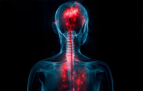
One of the longest nerves in the body is known as the vagus nerve (VN). The VN is the 10th pair of cranial nerves that originates at the brain stem in the medulla oblongata. This nerve is part of the parasympathetic nervous system, which is a part of the ANS. Research suggests ear acupuncture can activate the VN.
When to Order Advanced Imaging
For musculoskeletal imaging, the most commonly ordered studies are plain film radiographs. Under certain circumstances, computerized tomography (CT) or magnetic resonance imaging (MRI) may be required to differentiate a simple strain from a more complex problem such as a tumor, infection or degenerative disorder. There are symptoms and signs that can signify an injury or disease that may require advanced imaging or a referral to a specialist. These may include severe unrelenting pain at night; marked weakness; significant loss of range of motion; claudication; and systemic symptoms.
The use of CT or MRI should be based on your clinical findings. When a diagnosis cannot be determined, the CT or MRI should be considered only if the results will affect your treatment plan. You also need to consider the fact that these special studies are more sensitive than specific, and you will often have false positives.
The general rule of thumb is that CT is preferred for evaluating the cortex, trabecular structure and fractures. MRIs, on the other hand, are preferred for assessing soft tissue, bone marrow, ligaments, muscles, tendons and fat. MRIs are also useful in evaluating internal derangements of joints, metastatic diseases and primary tumors of the soft tissue.
Non-complex injuries such as a sprain/strain generally do not facilitate the need for advanced imaging. However, when plain film radiographs are inconclusive and conservative treatment has not helped, a CT or MRI will provide further evaluation.
When deciding to order an MRI or CT scan, a CT might be preferred because of price. However, you must consider that CT will expose your patient to ionizing radiation. A CT is also limited to scans of the axial plane, whereas MRI has the ability to image directly in a variety of planes without reconstruction. MRIs with the contrast agent Gd-DTPA (gadolinium diethylene-triamine penta-acetic acid) provide both physiologic and anatomic information. MRIs also have fewer occurrences of false negative results than CT scans.
When ordering advance imaging, we must also consider contraindications for CT and MRI. A CT is contraindicated in pregnancy and for use on children unless appropriate. MRIs are contraindicated with patients that have cardiac pacemakers or other ferromagnetic materials in their bodies such as transplants or clips.
Below is a non-exclusive list of selected indications for ordering a CT or MRI.
| Indication | Study Usually Performed |
| Bone | |
| Fractures, trauma, deformity | CT |
| Stress, occult, or minimally displaced fractures | MRI |
| Bone marrow (including lymphoma, myeloma) | MRI |
| Soft Tissues/Tumors and Masses | |
| Benign (bone) | CT |
| Benign (soft tissue) | MRI |
| Malignant (soft tissue or bone) | MRI |
| Metastases | MRI or contrast MRI, bone scan |
| Metastases, lung | CT |
| Hematoma | |
| Hematoma, bleeding into tissue | MRI |
| Epidural hematoma | MRI (CT if patient is traumatized) |
| Hip | |
| Avascular necrosis | MRI |
| Osteonecrosis | MRI |
| Transient osteoporosis | MRI |
| Infection, Inflammation, Abscesses, Osteomyelitis | MRI or contrast MRI |
| Intra-Articular | |
| Intra-articular structures | MR arthrography |
| Loose bodies in a joint | CT arthrography or MR arthrography |
| Spine | |
| Cauda equina syndrome | Emergent MRI |
| Degenerative disk disease | MRI or CT |
| Herniated disk, spinal stenosis | CT or MRI (possibly with myelography) |
| Low back pain w/ neurological signs | MRI or CT |
| Spondylolisthesis | Plain x-ray films are best |
| Labrum | |
| Tears and degeneration | MRI, MR arthrography |
| Rotator Cuff | |
| Full thickness tear | Arthrography, MRI |
| Partial thickness tear | MRI with contrast; MR arthrography preferred to arthrography alone and is better than conventional MRI |
| Shoulder impingement syndrome | MRI, MR arthrography, or plain x-ray films |
| Meniscus | |
| Meniscal injuries | MRI |
References
- Bluemka DA, Zerhouni EA. MRI of avascular necrosis of bone. Top Magn Reson Imaging 1996;8:231-246.
- Boegard T. Radiography and bone scintigraphy in OA of the knee comparison with MR imaging. Acta Radiol Suppl 1998;418:7-37.
- Boegard T, Rudling O, Petersson IF, et al. Correlation between radiographically diagnosed osteophytes and magnetic resonance detected cartilage defects in the patellofemoral joint. Ann Rheum Dis 1998;57:395-400.
- DePaulis F, Cacchio A, Michelini O, et al. Sports injuries in the pelvis and hip: diagnostic imaging. Eur J Radiology 1998;27(suppl 1):S49-S59.
- Ryan PJ, Reddy K, Fleetcroft J. A prospective comparison of clinical examination, MRI, bone SPECT and arthroscopy to detect meniscal tears. Clin Nucl Med 1998;23:803-806.
More reference are available upon request.


