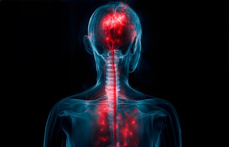
One of the longest nerves in the body is known as the vagus nerve (VN). The VN is the 10th pair of cranial nerves that originates at the brain stem in the medulla oblongata. This nerve is part of the parasympathetic nervous system, which is a part of the ANS. Research suggests ear acupuncture can activate the VN.
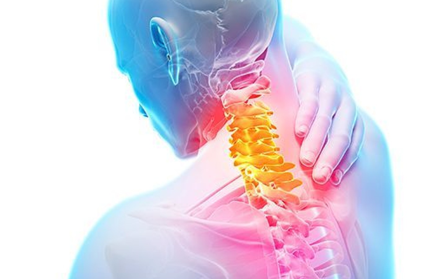
Understanding Cervical Compression
When evaluating the neck, there are any number of orthopedic tests to be considered. In my experience, most examiners check a neck complaint with just a simple Y-axis compression - commonly performed by having the patient tilt the head back slightly while the examiner applies downward pressure on the vertex – then note if there is pain. However, there are a number of other maneuvers that can be pursued and interpreted to give you a better understanding of the nature of the patient's complaints. I have seen these test names get used interchangeably, but they are unique and specific tests.
Cervical foraminal compression: Note the location of any discomfort with rotation of the head and return to neutral. Pain on the opposite side of rotation suggests muscular strain, while pain on the side of rotation suggests facet or nerve root involvement. Gently and gradually exert downward pressure to reproduce the pain. If pain is produced then turn the head to that side and perform the maneuver again. Reproduction of pain is indicative of foraminal encroachment. Radicular pain would then direct more thorough exam to the indicated neurologic level.
Jackson's Compression: Again with the patient seated have them rotate side to side and localize the complaint. Pain on the opposite side of rotation suggests muscular strain, while pain on the side of rotation suggests facet or nerve root involvement. Flex the head laterally (ear to the shoulder) and hold the position while exerting gentle pressure down on the head. The test is positive if the localized pain radiates down the arm. Note the nerve level and examine accordingly.

Maximum Cervical Compression: Patient seated with head in neutral. The head is actively rotated to the side of the complaint and then extends (lift the chin up). Increased pain on that side indicates nerve root or facet involvement – pain on the other side suggests muscular strain. A variation of this test may involve the same movements, but having the patient flex the head (look down) while rotating.
Spurling's: This test is a combination of some of the other maneuvers. Again have the patient sit and rotate the head – localize the pain. Laterally flex the head and gently press down to reproduce the pain. From the laterally flexed position extend the head as far back as the patient can tolerate – localized pain at this time suggests facet, radicular pain would indicate nerve root. Return to neutral. The well-known modification to this test is to then perform a quick, vertical blow down on top of the head – this impulse will fire off any pain generators including disc disease and cervical spondylosis.
Similar tests – slightly different moves to identify the pain vector. Make sure you know which one you are doing to properly document the findings in your records. Physical examination is a necessary part of a patient evaluation, and the standard of care requires accurate documentation. Certainly, there are other tests, signs and observations to be made in individual cases. The more information you have to confirm your findings, the more secure you are in your diagnosis and treatment protocol. Understanding and applying the different versions of cervical compression can only improve your examination findings and the specificity of your care. This extra documentation also can help make the difference if you must justify your diagnosis to an insurer or third party. Take the extra few seconds to add these tests into your exam routine - they will serve you well.
Resources
- Evans RC. Illustrated Essentials in Orthopedic Physical Assessment, 1994; Missouri: Mosby.
- Hoppenfeld S. Physical Examination of the Spine and Extremities, 1976; California: Appleton & Lange.


