Regarding patient care, I worry about inaction by the patient and lack of progress: if my patients are not putting into action something I have recommended to change, or if they are, but the symptoms are still not changing The most common things I recommend are eating changes, posture awareness, movement to stretch, strengthen or reawaken the nervous system, and improving sleep quality.
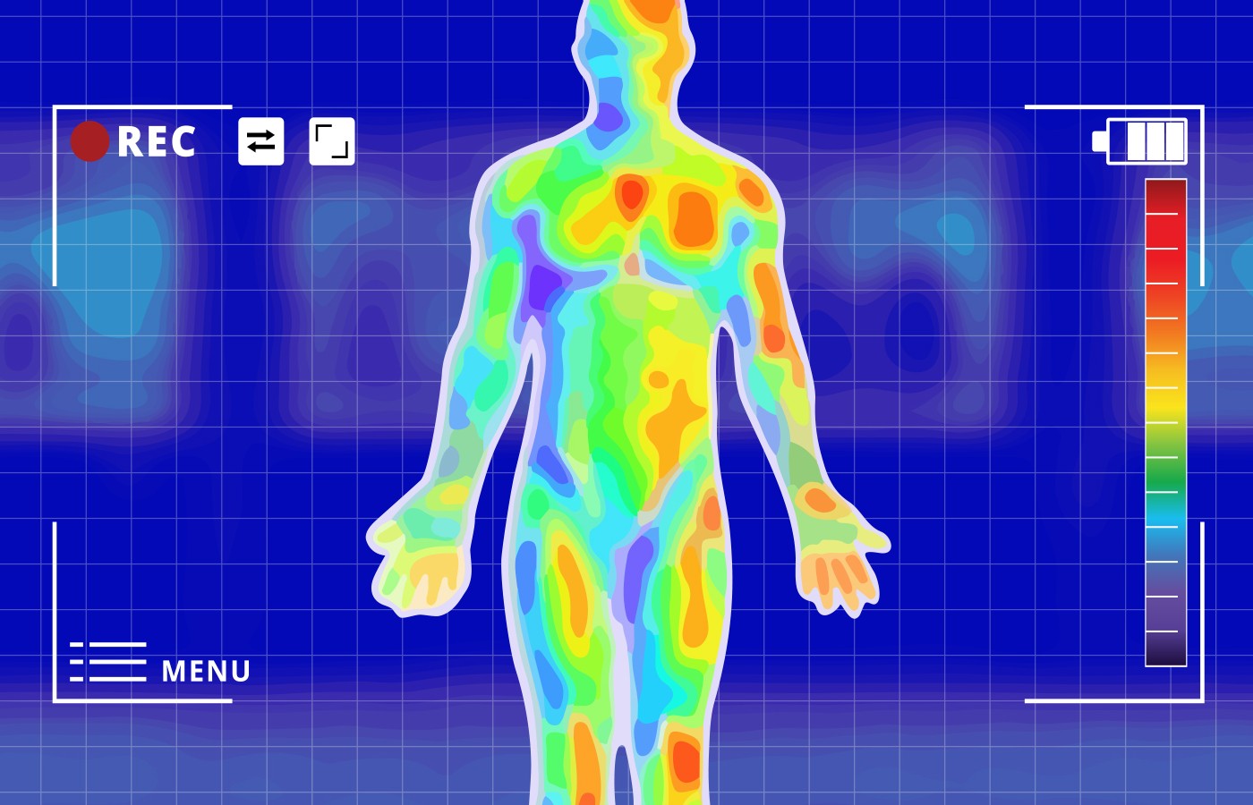
I’ve recently started using a new handheld thermography device that provides immediate thermal images and shows me (and my patient) particular areas of higher and lower temperatures. Musculoskeletal thermography (MT, also known as infrared thermography or thermal imaging), is a non-invasive tool that uses infrared cameras to detect and visualize variations in skin temperature.
This information is valuable on a visit-to-visit basis and helps me make treatment choices. For example, the infrared images have shown me remote areas from the chief complaint with increased or decreased temperature that I missed during my functional examinations. Thermal images can show correlations to old traumas, especially old ankle injuries, and patients see the overcompensation effects in other areas, or the opposite side of the body, related to current complaints.
Thermography is helping me demonstrate to the patient where adverse loads are being placed in their body and is putting more into action on their own – think gamification!
Practice Application
Here are some of the top uses for thermal imaging in musculoskeletal health:
Injury assessment: Infrared thermography can help assess the severity and extent of injuries by detecting areas of inflammation, muscle strains, ligament sprains, and other soft-tissue damage. Areas of increased temperature surrounded by a normal temperature area may be related to increased blood flow or friction (heat producing) related to densification of fascia.
On the flip side, it has been particularly useful in identifying cooler areas that correlate to neurological deficiencies and may respond to light and heat therapy. These images are guiding my palpation to remote areas, and I am picking up more trigger points and tender points that were not apparent through other digital examination methods.
Pain evaluation: With the patient sitting next to me, together we view black-and-white photos become colorful, thermal images, and we can identify the source and intensity of pain in their musculoskeletal conditions. In real time, I provide objective evidence of abnormal temperature patterns associated with conditions such as arthritis, tendinitis, myofascial pain syndrome, and nerve entrapment syndromes.
Monitoring treatment & rehabilitation: So far, my favorite part of using thermography is to monitor the progress of treatments and therapies. By comparing thermal images taken at different stages of treatment (intersession, at the beginning of each session, etc.), it becomes possible to assess changes in inflammation, blood flow and tissue healing.
For example, treating a higher-temperature “spot” with laser and watching the temperatures normalize demonstrates we are accomplishing our goals. The same is true of cooler areas.
Sports medicine and equine medicine: Infrared thermography is widely used in sports medicine and veterinary clinics to detect and prevent injuries. It can help identify areas of increased stress, muscle imbalances and areas at risk of injury. This information can be utilized to modify training programs, prevent further damage and optimize performance.
Occupational health and ergonomics: Thermography is valuable in assessing work-related musculoskeletal disorders (WMSDs) and evaluating ergonomic conditions in the workplace. It can aid in identifying areas of increased stress, poor posture and improper biomechanics, all of which can contribute to the development of WMSDs.
Assessment of vascular disorders: Musculoskeletal thermography can assist in evaluating circulatory disorders affecting the extremities. By assessing skin temperature patterns, it can help detect peripheral vascular diseases such as deep-vein thrombosis, peripheral artery disease and Raynaud’s disease.
Research and education: This technology provides valuable visual data for analysis, and helps in understanding the mechanisms underlying various conditions and injuries.
Clinical Takeaway
In the less than 2-5 minutes it takes to capture and explain thermal images, it is guiding my treatment recommendations and uncovering new ways to boost the nervous system and regulate blood flow. I have less “overwhelm” in where to start my treatment process and what therapy to apply. It makes me more decisive and immediately stimulates greater patient “gamification,” which provides a boost in patient compliance.
Thermography continues to bring tremendous value to my practice, with very little logistical interruption and immediate impact to patient care and education.