Cupping therapy is widely employed in traditional East Asian medicine for musculoskeletal pain, stagnation and systemic conditions. While the formation of erythema and ecchymosis is an anticipated skin response, the appearance of post-treatment vesicles remains less discussed in clinical literature.
This article presents a case study of vesicle formation following cupping therapy, explores underlying pathophysiology, and offers management guidelines to support practitioners in addressing this phenomenon safely and effectively.
Cupping therapy, a hallmark modality within traditional Chinese medicine (TCM), has been practiced for centuries to invigorate qi and blood circulation, expel pathogenic factors and relieve pain. With its growing global popularity, practitioners increasingly encounter diverse patient presentations and tissue responses, necessitating greater clinical discernment.
Although local skin discoloration post-cupping is a well-recognized outcome, the development of fluid-filled vesicles is less frequently addressed in professional dialogue. Understanding the mechanisms, risk factors, and appropriate management of this response is critical for ensuring patient safety and optimizing therapeutic outcomes.
Case Presentation
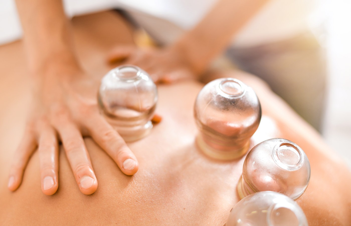
A 37-year-old female fitness professional presented for cupping therapy to address chronic thoracic and trapezius tension. Stationary silicone cups were applied bilaterally along the Bladder Meridian (BL 13 – BL 17 region) using moderate-to-strong negative pressure for a duration of 12 minutes. Treatment was well-tolerated, with expected post-treatment ecchymosis and erythema.
Within 24 hours, the patient noted the appearance of multiple small, clear vesicles (1-4 mm) over several cupped regions. She reported no pain, pruritus or signs of infection. The vesicles resolved spontaneously over five days with conservative skin care and without sequelae.
Pathophysiology
From a biomedical perspective, cupping induces local capillary rupture, increased vascular permeability and interstitial fluid accumulation. Excessive negative pressure or prolonged application can create mechanical separation between the epidermal and dermal layers, resulting in vesicle formation. The clear fluid typically consists of plasma, lymphatic fluid and interstitial components.
Within the TCM framework, vesicles may reflect the successful externalization of dampness or toxic heat in cases of localized pathogenic accumulation. However, they may also indicate that the intensity of the intervention exceeded the patient’s constitutional tolerance or the local tissue’s capacity for adaptive response.
Clinical Implications
Patient Communication: Practitioners should provide pre-treatment counseling on possible skin responses, including ecchymosis, petechiae and, less commonly, vesicles. Framing these outcomes within both biomedical and TCM perspectives can improve patient confidence and mitigate anxiety if vesicles develop.
Risk Factors and Contraindications: Thin, sensitive or aged skin; high negative pressure or prolonged retention time; recent exfoliation, sunburn or dermatologic conditions (e.g., eczema, psoriasis); immunocompromised or corticosteroid-dependent patients.
Management Strategies: Advise patients not to rupture vesicles; maintain skin integrity. Recommend gentle cleansing and, if needed, application of a non-occlusive antibacterial ointment. Encourage wearing loose, breathable clothing over the area. Monitor for signs of secondary infection (erythema, warmth, pain, purulence). Adjust subsequent treatments by reducing pressure or time and considering moving cupping techniques.
Discussion
Vesicle formation serves as an important marker of tissue response and requires nuanced interpretation. While generally benign, it underscores the need for individualized treatment planning. Practitioners must balance therapeutic vigor with tissue resilience, particularly when working with patients unfamiliar with cupping or those presenting with predisposing factors.
It is essential to recognize that stronger stimulation does not necessarily equate to better outcomes. Rather, the skilled practitioner modulates intensity to match the patient’s presentation, constitution and treatment goals.
Vesicle formation after cupping is a rare but noteworthy clinical event. With proper patient education, risk assessment and management, practitioners can prevent complications and maintain the therapeutic alliance. As cupping continues to expand in clinical practice, ongoing dialogue and research are encouraged to further elucidate best practices in managing post-cupping skin responses.