For more than 2,500 years, traditional Chinese medicine practitioners have formulated various theories on the meridian system and mechanisms as the basis of acupuncture. Practitioners and researchers have been searching for the replicable anatomical structures of meridians. There are researchers who don’t believe there are meridian structures other than nerves and blood vessels.1
What is the anatomical morphological basis for meridians? One study, based on anatomically dissected human specimens, identified extracellular matrix and fascia, along with vessel-nerve bundles, as important parts of the anatomic substrate of acupuncture meridians, with muscles, tendons and ligaments that follow the meridian course.2 This finding differs from the theory that acupuncture points are primarily located along the nervous channels.3
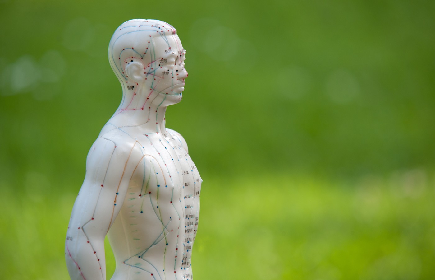
New York acupuncturist Ning Ma believes, “The meridian is an independent human body structure system that is not classified according to the modern anatomical system.” Rather, the meridian is like a passageway that runs through the interstitial spaces, is wrapped by fascia, and is accompanied by blood vessels and nerves.4
Ma reconstructed all the meridians in his book, published in May 2023. He clearly and meticulously describes the body surface landmarks, bones, muscles, ligaments, blood vessels, nerves, fascia, and body cavities; as well as the connection and transformation between various tissue structures along this passageway.
Part of the circulation of the spleen meridian of foot taiyin is described as follows in Ma’s book:
“starts from the end of the great toe at the base of the distal phalanx, runs along the tissue space around the flexor hallucis longus tendon; around the metatarsophalangeal joint, goes into the tissue space around the abductor hallucis and adductor hallucis muscles; after navicular tuberosity of the medial malleolus, distributed from the plantar to the dorsum of the foot along the medial malleolus artery; then it runs in the posterior fascial sheath of the calf, transition through the posterior tibialis muscle to the tissue space around the sartorius muscle; it migrates between the transversus abdominis and the internal fascia, enter the diaphragmatic splenic ligament through the diaphragm and connects with the spleen. The marked body surface landmarks are: the lower border of the abductor hallucis muscle is the border of red and white flesh, and the medial malleolus scaphoid tuberosity is the nuclear bone.”4
The description of meridians in traditional acupuncture textbooks can easily give the impression meridians are two-dimensional paths that connect acupuncture points through the body surface. In contrast, the meridians restored by Ma contain the skin, muscles, tendons, and veins, including three-dimensional channels with layers and depths. This echoes the anatomical morphological basis for meridians observed by Maurer.4
Dr. Helene Langevin found that “needle grasp” caused by connective tissue / fascia winding around the needle is the biomechanical component of de qi.5 If we can further observe the structural coherence and functional interaction of these passageways, we may be able to further verify the advisability of Ma’s meridian model.
Prior to this, learning types of meridian anatomical models like Ma’s may help us to more accurately select the depth and angle of needling, choose the combination of far and near points, and match the needling method with different tissues of the skin, muscles, tendons, bones, and veins to achieve the desired effects.
With reference to this type of meridian anatomical model, the acupoints and meridians in the acupuncture medical records will present a three-dimensional description that is highly compatible with the modern anatomical system, making it easier to achieve clinical communication and improvements on evidence-based medicine in acupuncture.
References
- Zhang S-J. [Practice and diversion of acupuncture-moxibustion scientization in modern times: taking ZHU Lian as the center.] Zhongguo Zhen Jiu (Chinese Acupuncture & Moxibustion), October 2014;34(10):1009-15.
- Maurer NN, et al. Anatomical evidence of acupuncture meridians in the human extracellular matrix: results from a macroscopic and microscopic interdisciplinary multicentre study on human extracellular matrix. Evid Based Complement Alternat Med, 2019 Mar 21;2019:6976892.
- Zu L. [New Acupuncture]. Nan Ning (Publishing City): Guang Xi Ren Min Chu Ban She (Guangxi People’s Publishing House),1980.
- Ma N. [Anatomy of Channels and Collaterals]. Jin Nan (Publishing City): Shan Dong Ke Xue Ji Shu Chu Ban She (Shandong Science and Technology Publishing House), 2023.
- Langevin HM, et al. Mechanical signaling through connective tissue: a mechanism for the therapeutic effect of acupuncture. FASEB J, 2001;15(12):2275-2282.