Editor’s Note: The primary author of this article, Dr. Su, credits Dr. Arthur Yin Fan for his contributions to this article.
Last year, I participated in the 10th annual American Traditional Chinese Medicine Conference in Las Vegas. This was my first time attending such a high-level academic event, and it was truly eye-opening. One of the most profound takeaways was the extensive introduction of scalp acupuncture during the conference.
Over the two days, several professors shared their unique scalp acupuncture protocols. It sparked many reflections in my mind: Scalp acupuncture treatments have demonstrated clinical outcomes; what are the internal brain changes after treatment? What are the theoretical foundations of these methods?
During the conference, Professor Jason Hao’s introduction to scalp acupuncture was fundamentally based on Jiao’s scalp acupuncture technique, which integrates traditional acupuncture with modern neuroanatomical and neurophysiological principles. A region of the scalp extending from the vertex to the temporal area resembles an inverted, miniaturized version of the human body, divided into bilateral sensory and motor zones – with one-fifth representing the lower limbs, two-fifths the trunk and upper limbs, and two-fifths the head and face.
The anterior portion before bilateral sensory and motor zones is designated as the tremor zone, while the crown represents the foot sensory area, with three additional language regions.
Professor Hao’s needle insertion technique is distinctive and swift, involving multiple rapid rotations of the needle beneath the aponeurosis at a speed of 200 rotations per minute. He asserted that this method of scalp acupuncture is effective in treating central nervous system disorders such as stroke-induced hemiplegia and aphasia.
I have also employed this technique in my practice, and my preliminary observation suggests it is generally painless and well-accepted by patients. However, the number of cases remains limited, and further observation of its therapeutic efficacy is required.
Professor Wei Liu introduced the basic Liu's scalp acupuncture protocol, primarily aimed at treating pain disorders. Conference materials revealed that this method is based on Fang’s scalp acupuncture holistic cranial positioning, with Baihui (Du 20) corresponding to the body acupuncture point Mingmen (Du 4). Professor Liu explained that this technique is his interpretation of the Huangdi Neijing (Yellow Emperor’s Inner Classis) and Nanjing (The Classic of Difficult Issues). He emphasized that mastering acupuncture requires a thorough study of the Ling Shu section of Neijing.
During the conference, Professor Liu demonstrated his method by applying scalp acupuncture combined with manual therapy for patients with lower back pain, which he termed the "Liu’s Kite Needle Technique."
Professor Xiping Zhou emphasized the equal importance of both massage techniques and acupuncture in clinical practice. In this session, Professor Zhou demonstrated six distinct rolling massage techniques. I recalled that Professor Zhou was also skilled in scalp acupuncture. Previously, I had seen online his introduction of Zhou's technique, which centers on Baihui, with needle insertions extending to a rhombic area defined by the four directions of the Sishencong points. He asserted that this method yields significant clinical results.
Additionally, it occurred to me that in Professor Guanyuan Jin’s 2023 lecture on scalp acupuncture and reflection points, he discussed how scalp acupuncture stimulation improves cerebral blood flow. He also highlighted the effectiveness of the simplified Jin’s four major zones (vertex, frontal, temporal, and occipital), along with auricular acupuncture, in achieving favorable clinical outcomes.
During the conference, I discussed the different scalp acupuncture’s benefits with Dr. Arthur Yin Fan, who explained the objective changes in brain function, particularly with the cerebral cortex, before and after scalp acupuncture. He recommended further exploration of the literature and suggested I could compile a review on this subject. After the conference, I revisited Dr. Fan's previous Zoom lecture organized by the American Traditional Chinese Medicine Association (ATCMA),1 in which he introduced the concepts of the "brain-specified acupuncture points (acupoints)" and the "brain-specified acupuncture zones (acuzones)."
Dr. Fan emphasized the unique role of the brain as a vital organ, noting that it possesses specific acupuncture points located in areas with dense head muscles, such as the forehead, temporal region, occipital area, upper cervical region, and the eye socket. He identified three distinct stimulation zones for scalp acupuncture.
The first zone, the "brain-specified acupoint" zone, is divided into two areas: 1A, which includes the area around the eyebrows, eye socket, lower jaw, and frontal muscles, extending to the temporal muscles near points like Taiyang, Xuánluo, and Shùigu, as well as the corresponding skin and connective tissues; and 1B, the occipital-cervical transition zone, which includes the occipital muscles, upper cervical muscles, and corresponding skin and connective tissue.
This first zone generally does not experience pain or tenderness during a normal physiological time; however, during the disorders of neurological conditions or episodes, the muscles in the head and neck at zone 1A and 1B may experience varying degrees of swelling, tenderness, and sensitivity to touch, making this area particularly significant as the "brain-specified acupuncture points."
The second zone encompasses the forehead along the hairline, while the third zone refers to the crown of the head, which forms a rhombus-shaped region extending one inch from the four points of Sishencong, and the area Naokong (GB 19) to Naohu (Du 17). The second and third zones generally do not show significant pain, tenderness, or tissue swelling before or after the onset of neurological conditions, which is why they are not considered part of the "brain-specified acupoint" zone, although they are still related to adjusting the brain functions.
The general principle of acupuncture stimulation in these regions involves the activation and regulation of multiple layers and multiple nuclei in the brain, especially within the sensory and motor areas. For example, stimulation within the trigeminal nerve distribution zone primarily regulates several regions of the brain, while stimulation in the occipital nerve (primarily C2) distribution zone mainly affects the cerebellum and brainstem.
Notably, acupuncture at different scalp locations does not exhibit a distinct specificity – needle insertion at a particular site does not only affect the corresponding sensory or motor cortex directly beneath the needle-stimulated scalp area, but instead activates or inhibits multiple cortical and subcortical areas. The overlap between the two neural distribution zones (trigeminal nerve and C2 nerve) in functional regulation is significant. (For a more detailed discussion, please refer to the ATCMA 2024 online public lecture.)
I read another article about Jiao’s scalp acupuncture, which was published by Dr. Mingzhu Ye and Dr. Arthur Yin Fan. The conclusion is that scientific evidence hasn’t confirmed Jiao’s hypothesis, nor has that hypothesis followed from the development of neuroscience. Jiao's insistence on his hypothesis seriously hindered the research and development of scalp acupuncture therapy.
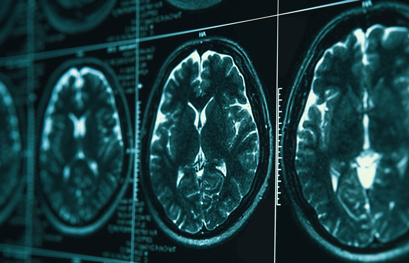
Clinical trials and experimental studies on acupuncture, especially using an fMRI, are warranted to evaluate scalp acupuncture’s therapeutic value and to identify the functional changes of various parts of the brain that acupuncture on the scalp can cause.2
To explore the mechanisms of scalp acupuncture, I further reviewed objective studies on brain function changes following scalp and body acupuncture treatments. Using functional MRI (fMRI) as objective evidence, it was found that several clinical research studies and meta-analyses have provided valuable insights.
Acupuncture has been recommended by the World Health Organization as an alternative and adjunctive strategy for stroke treatment. A substantial body of research indicates that acupuncture has a positive therapeutic effect on stroke patients, aiding in motor recovery, alleviating dysphagia, reducing shoulder pain, improving cognitive impairments, alleviating depression, and addressing other symptoms.
Evidence suggests that the mechanisms underlying acupuncture's effectiveness in stroke treatment primarily include promoting neurogenesis and cell proliferation, regulating cerebral blood flow in ischemic areas, and exerting anti-inflammatory and anti-apoptotic effects.3
fMRI is a non-invasive imaging technique with high temporal and spatial resolution, which measures neuronal activity by monitoring hemodynamic responses.4 Increasingly, acupuncture research is incorporating fMRI technology to explore the neural effects associated with treatment responses, aiming to better understand the underlying mechanisms of acupuncture and evaluate its therapeutic efficacy.5
Ma, et al.,6 conducted a meta-analysis that examined changes in brain fMRI following acupuncture treatment in patients with mild cognitive impairment (MCI). Although this study did not specifically focus on scalp acupuncture, the use of fMRI as objective evidence offers valuable insights and considerations. The meta-analysis included seven studies that met the inclusion criteria, with a treatment group of 94 patients and a control group of 112 patients. All studies utilized the Regional Homogeneity (ReHo) method for analysis. The fMRI experimental design included six task-related studies and one resting-state study.
The results indicated that acupuncture treatment led to enhanced activity in the right insula, left anterior cingulate/precuneus, right thalamus, right middle frontal gyrus, right posterior cingulate/precuneus, and right middle temporal gyrus in MCI patients.
Further analysis through randomized, controlled trials (RCTs) and longitudinal studies revealed a significant increase in ReHo values in two brain regions (left anterior cingulate/precuneus and right insula) following acupuncture. Compared to healthy controls, MCI patients exhibited stronger activity in the right parahippocampal gyrus after acupuncture treatment. Meta-regression analysis showed a significant correlation between the ReHo index of the right anterior thalamus projection and the post-acupuncture MMSE scores in all MCI patients.
Li, et al.,7 conducted a meta-analysis on brain MRI changes following acupuncture treatment in patients with aphasia. After reviewing 885 articles, 16 studies were ultimately included. Regarding acupuncture types, 10 studies employed manual acupuncture, while five utilized electroacupuncture. Body acupuncture (10 studies), scalp acupuncture (seven studies), and tongue acupuncture (eight studies) were used to treat post-stroke aphasia.
Results suggested the mechanism of acupuncture may be related to the activation and functional connectivity of language-related brain areas, such as the left inferior temporal gyrus, supramarginal gyrus, and Broca's and Wernicke's areas, as well as the frontal gyrus and inferior frontal gyrus. However, these studies are still in the preliminary stages. Large-scale, multicenter RCTs are needed to validate the existing evidence and to further explore the neuroimaging mechanisms underlying acupuncture effects.
Editor’s Note: Part 2 of this article reviews additional research on the relationship between scalp acupuncture and brain activation in stroke patients.
References
- Dr. Arthur Fan. Evidence-based Scalp Acupuncture. ATCMA-TCMAAA online Seminar,02042024.
- Ye MZ, Fan AY, Alemi SF. Questioning the fundamentals of Jiao’s scalp acupuncture: point-positioning based on the hypothesis of the cerebral cortex’s functional zone. Altern Ther Health Med, 2023 Oct;29(7):242-251.
- Chavez LM, Huang SS, MacDonald I, et al. Mechanisms of acupuncture therapy in ischemic stroke rehabilitation: a literature review of basic studies. Int J Mol Sci, 2017 Oct 28;18(11):2270.
- Glover GH. Overview of functional magnetic resonance imaging. Neurosurg Clin N Am, 2011 Apr;22(2):133-9, vi.
- Zhang J, Li Z, Li Z, et al. Progress of acupuncture therapy in diseases based on magnetic resonance image studies: a literature review. Front Hum Neurosci, 2021 Aug 20;15:694919.
- Ma SQ, Huang HP, Zheng Z, et al. Effect of acupuncture on brain regions modulation of mild cognitive impairment: a meta-analysis of functional magnetic resonance imaging studies. Front Aging Neurosci, 2022 Sep 23;14:914049.
- Li B, Deng S, Sang B, et al. Revealing the neuroimaging mechanism of acupuncture for poststroke aphasia: a systematic review. Neural Plast, 2022 Apr 21:2022:5635596.